Tumor Growth and Dissection
•Sedentary and exercised cohorts were administered flank injections with MTLn3 rat mammary adenocarcinoma cell line
•Tumor growth monitored and voluntary wheel-running recorded
•21 days post-injection or when tumors grew to three centimeters, rats were euthanized using sodium pentobarbital
•Tumor growth monitored and voluntary wheel-running recorded
•21 days post-injection or when tumors grew to three centimeters, rats were euthanized using sodium pentobarbital
Immunohistochemistry
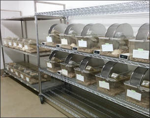
•Spleen and tumor tissue was excised from rats and stored in cryomolds with OCT compound
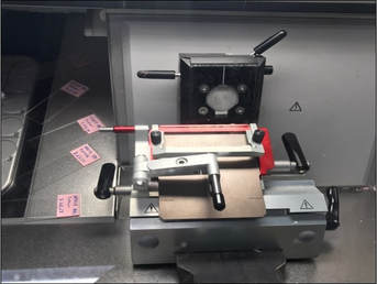
•Samples cryosectioned to 4 microns at -20°C and placed onto poly-D-lysine coated slides
•Tissues were fixed in acetone
•Washed with TBS (0.05% Tween 20 in PBS)
•Made permeable with 0.1% Triton-X 100 in PBS
•Blocked using 1% NGS (goat serum) in PBS
•Samples were stained with antibodies for CD25 and Foxp3 and held at 4°C until washed with TBS
•Hoechst dye was applied to all tissue samples to visualize cell nucleus through DAPI filter
•Antigen retrieval attempted (Sudan Black)
Visualization
|
•Samples were visualized using fluorescence microscopy
•2 fields of view randomly selected •Observed at 40x magnification |
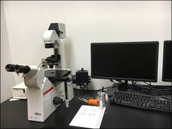
This is an image of the fluorescent microscope, which is the technology we used to collect images of the regulatory T cells
|
