Results
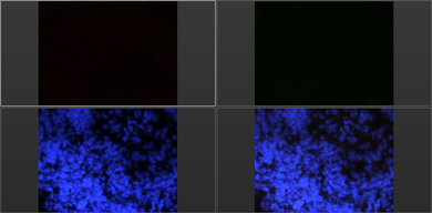
Negative control picture, with DAPI filter on bottom left that shows the cell's nuclei. Top left is where CD25 antibodies would be stained, and top right is where Foxp3 would be fluorescing. Bottom right is an overlay of three previous pictures.
|
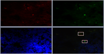
Same labels as previous picture. If a cell has a nucleus and fluoresces under the PE filter and FITC filter, then it is a Treg cell. There are two Treg cells in this field of view, and are boxed in white.
|
|
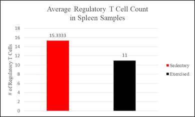
This is a graph comparing the average regulatory T cell count in spleen samples, with a P-value of 0.527. This data has no statistical significance when alpha is 0.05.
|
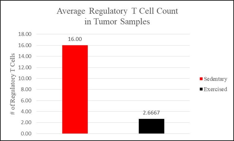
This is a graph comparing the average regulatory T cell count in tumor samples, with a P-value of 0.300. This data has no statistical significance when alpha is 0.05.
|
|
|
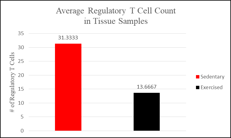
This is a graph comparing the average regulatory T cell count in all tissue samples, with a P-value of 0.347. This data has no statistical significance when alpha is 0.05.
|
Discussion
Treg cells were counted using 2 random fields of view per positive control sample. The positive CD25 cells and positive Foxp3 cells that have nuclei are regulatory T cells. The negative control, as shown in Figure 2, had stained nuclei, but did not fluoresce Foxp3 or CD25 antibodies as they were not put on the slide. The negative control group was to show that the procedure followed caused Treg cells to fluoresce, and the cells did not fluoresce on their own. Since the procedure did work, the negative control group did not fluoresce. The positive control group that were stained with antibodies, fluoresced which shows that the procedure worked in this area too.
None of our quantitative data supports our hypothesis as they are lacking statistical significance. When α = 0.05, we fail to reject the null hypothesis as none of the P-values were less than α. However, the data is trending towards a decrease of Treg cells in exercising animals
Limitations that were present in this study include having a novel protocol, limited population size, change in cancer cell line, and lack of previous research to compare data to. The protocol was limited as it was made specifically for this experiment, and there is no other research using a similar protocol. A possible reason as to why the data was not statistically significant may have been due to the small population size, with only 3 rats per cohort. Also, this protocol was the first successful Foxp3 IHC (immunohistochemistry) protocol, and could be adjusted for future studies. The lack of CD4 antibody would help in differentiating Breg and Treg cells, as both cell types fluoresce Foxp3+ and CD25+. The cell line was changed from MATB3 to MTLn3. MATB3 is a less aggressive cancer cell line - which would let researchers collect more data – and would therefore be more comparable to progression of cancer in humans.
